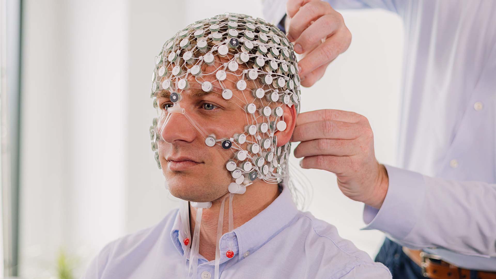
Saline or gel electrodes, depending on the system and configuration. Explore the brain's electrical activity illustrated on the model, then hover over an electrode to display its EEG signal.
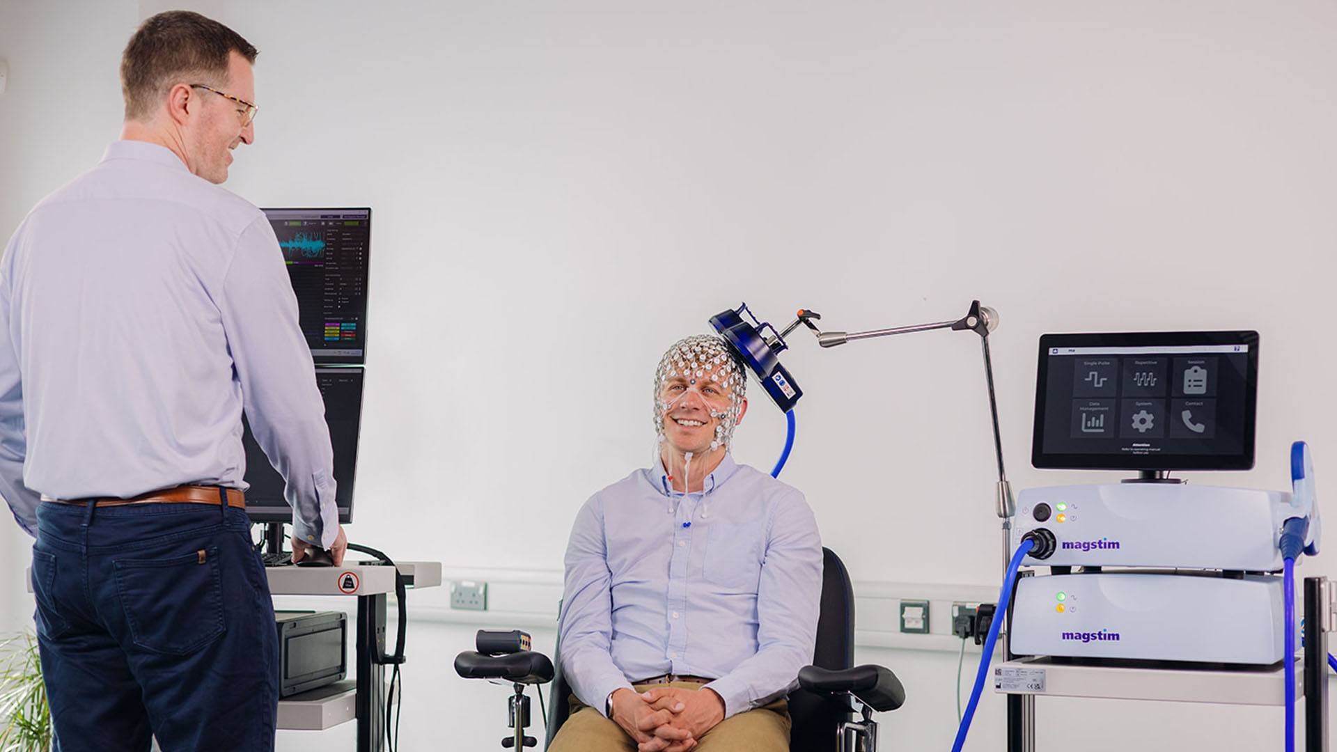
Educational animation · simulated signals, no real acquisitionThe coil and EEG are shown together. EEG traces are illustrative: they do not predict stimulation responses or artefacts.
Explore neuromodulation
Click the coil to pick it up, then click again to place it. Drag elsewhere to rotate the model.
Blue: magnetic field · orange-red: illustrative local activation
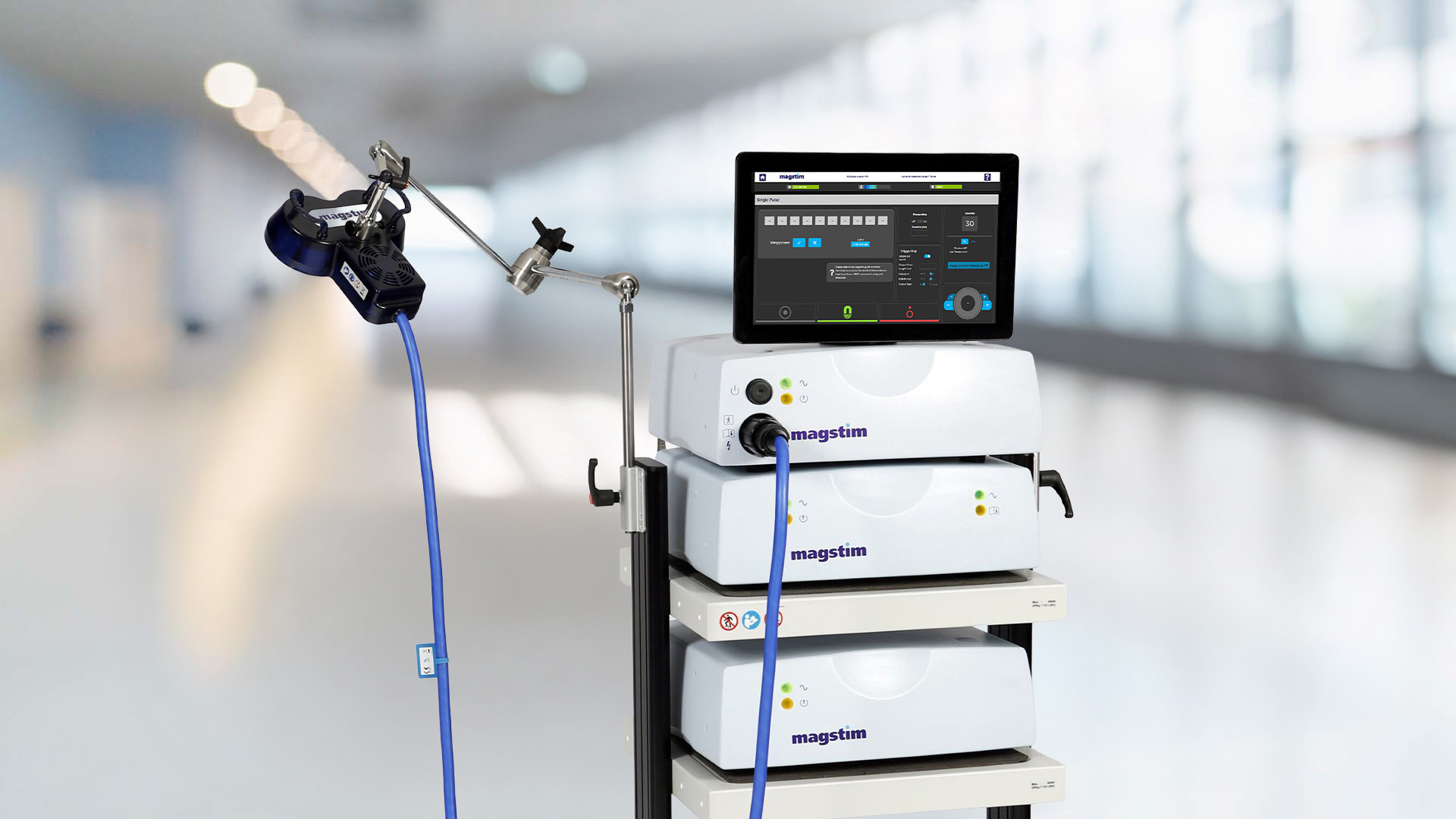
The changing magnetic field induces an electric field in tissue. Slowed, schematic animation, without dosage information or prediction of effects.Explore TMS equipment →
16 sources × 16 detectors
SourceDetector
Light diffuses from source to detector through a banana-shaped volume. Hover over an optode to explore simulated HbO and HbR changes.
Source-detector ≤ 3 cm · schematic depth, not guaranteed
Hold to rotate · Hover to explore · Hover over an electrode to view its signalOne finger to rotate · Tap an electrode to view its signal
Your curiosity leads the way.
Hover over the brain to discover its regions, or choose a technology: electrical activity with EEG, light and oxygenation with fNIRS.
A model to explore. Technologies to discover.
Model and sources
fNIRS: coloured light paths illustrate diffusion through tissue, not visible rays or exact photon trajectories. The scalp and skull are not represented anatomically; the cortex becomes translucent for the demonstration. Depth is capped at 3 cm in this simulation; this is not a device measurement specification. The 16 sources and 16 detectors do not represent 256 independent channels: only neighbouring pairs are illustrated. Principle: Understand the principle of fNIRS.
The EEG demonstration shows a schematic electrode arrangement around the cortex on a simplified scalp surface. Neurons and their connections are stylised and enlarged. Colours and traces are synthetic and represent neither real acquisition nor calculated source localisation.
Educational anatomical model. The exploded view separates structures for easier observation; it does not represent their natural positions.
Functional references: Johns Hopkins Medicine. Simplified explanations for introductory learning.
FROM SELECTION TO USE
Equipment advice
System sales
Integration and training
Technical support
01 / EEG, TMS & INTEGRATION
Record. Stimulate. Bring them together.
High-density EEG, magnetic stimulation or TMS–EEG integration: choose an individual system or a solution combining both. EGI EEG in France, Spain and Portugal; standalone TMS in France.
ACQUISITION01
High-density EEG
MagstimEGI
Equip your laboratory to record brain electrical activity. We help you choose the system, sensor nets and software for your recordings.
Choice of channel count and sensors
Configuration for your participants and environment
The EGI GES 500 and Magstim TMS systems bring together high-density EEG acquisition and magnetic stimulation, with a shared interface in Net Station 6.
Website language does not determine the sales country. Terms depend on the installation country and requested configuration.
EGI high-density EEG
France · Spain · Portugal. Exclusive EGI distribution in Spain and Portugal.
Standalone TMS
Standalone equipment sales: France only.
Standalone Faros
Standalone equipment sales: France only.
Eye tracking
Distribution throughout Europe.
fNIRS
France · Official NIRx representative in France.
MRI equipment
Distribution throughout Europe.
Integrated multimodal solution
Integrated multimodal solutions: projects worldwide, following Gaze Intelligence’s configuration review and approval.
Adding an accessory or several items does not by itself constitute a multimodal solution. Describe the modalities to synchronise and the scientific objective.